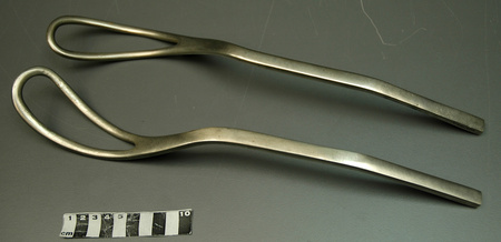
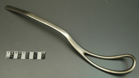
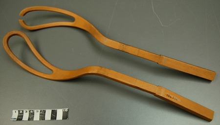
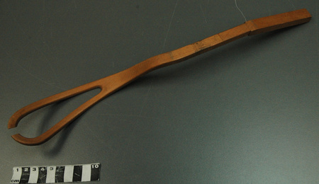
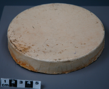
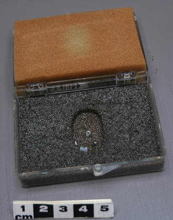
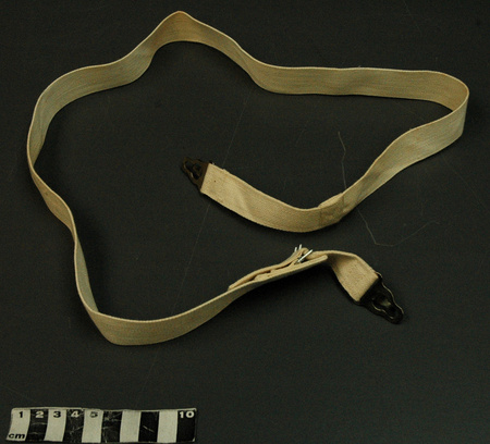
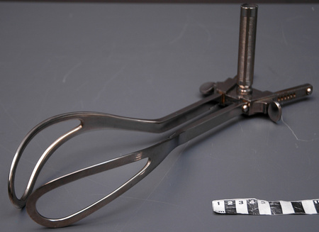
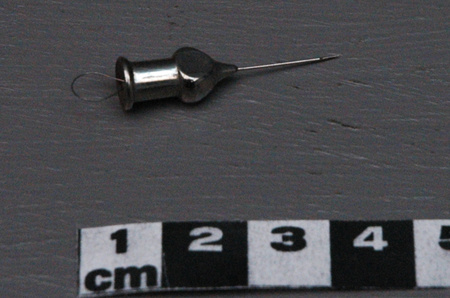
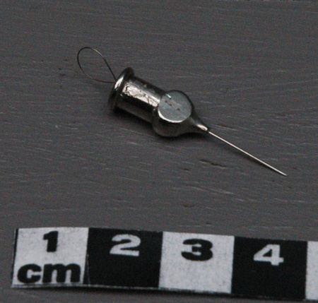
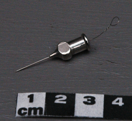
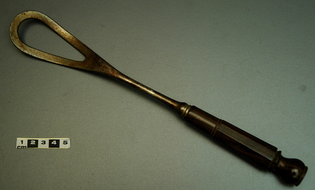
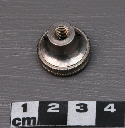
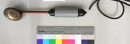
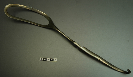
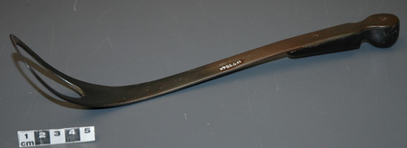
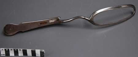
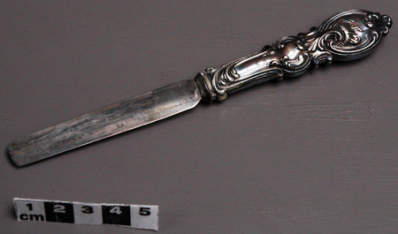
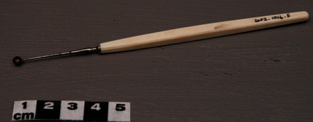
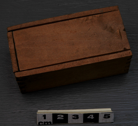
Blade
Use this image
Can I reuse this image without permission? Yes
Object images on the Ingenium Collection’s portal have the following Creative Commons license:
Copyright Ingenium / CC BY-NC-ND (Attribution-NonCommercial 4.0 International (CC BY-NC 4.0)
ATTRIBUTE THIS IMAGE
Ingenium,
2002.0618.002
Permalink:
Ingenium is releasing this image under the Creative Commons licensing framework, and encourages downloading and reuse for non-commercial purposes. Please acknowledge Ingenium and cite the artifact number.
DOWNLOAD IMAGEPURCHASE THIS IMAGE
This image is free for non-commercial use.
For commercial use, please consult our Reproduction Fees and contact us to purchase the image.
- OBJECT TYPE
- N/A
- DATE
- 1950–1965
- ARTIFACT NUMBER
- 2002.0618.002
- MANUFACTURER
- Schering
- MODEL
- Unknown
- LOCATION
- Unknown
More Information
General Information
- Serial #
- N/A
- Part Number
- 2
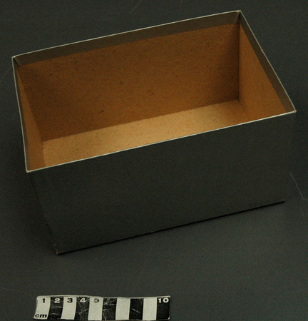
- Total Parts
- 4
- AKA
- N/A
- Patents
- N/A
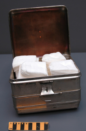
- General Description
- metal
Dimensions
Note: These reflect the general size for storage and are not necessarily representative of the object's true dimensions.
- Length
- N/A
- Width
- 1.0 cm
- Height
- N/A
- Thickness
- N/A
- Weight
- N/A
- Diameter
- 5.0 cm
- Volume
- N/A
Lexicon
- Group
- Medical Technology
- Category
- Radiology
- Sub-Category
- N/A
Manufacturer
- AKA
- Schering
- Country
- Unknown
- State/Province
- Unknown
- City
- Unknown
Context
- Country
- Canada
- State/Province
- Unknown
- Period
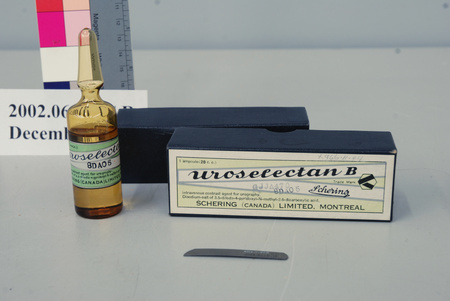
- This blade never used. This brand of contrast media probably available & used c. 1950- c. 1965, and possibly later.
- Canada
-
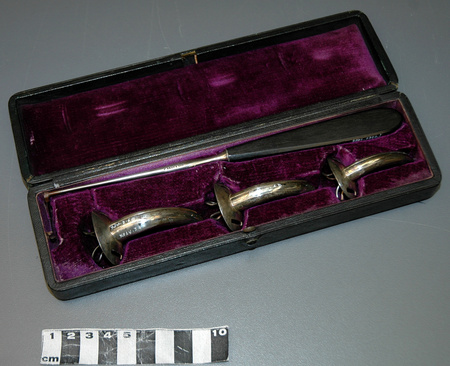
Part of a large collection of medical artifacts, archival material & trade literature transferred to CSTM in 2002 from the former History of Medicine Museum, Toronto ON. Supplied with brand of contrast media distributed in Canada by Schering (Canada) Ltd., Montreal, PQ. Originally donated to the History of Medicine Museum- Academy of Toronto c. 1966 by Dr. Walter Little Carruthers (1890-1968). [Ref. 2] - Function
-
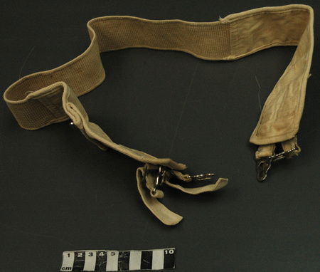
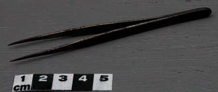
Used to remove the top of sealed glass ampoule, in order to access contents. - Technical
-
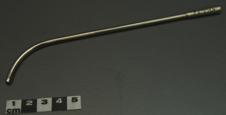
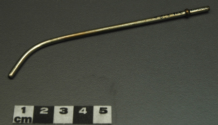
Urography (pyelography) is the X-ray examination used to view the kidneys, bladder and associated structures. Contrast media (dye) in administered intravenously, and a series of X-rays taken at intervals. Urography is used to investigate a range of problems including kidney pain and/or stones, blood in the urine, or a suspected obstruction or abnormality (congenital). [Ref. 6] - Area Notes
-
Unknown
Details
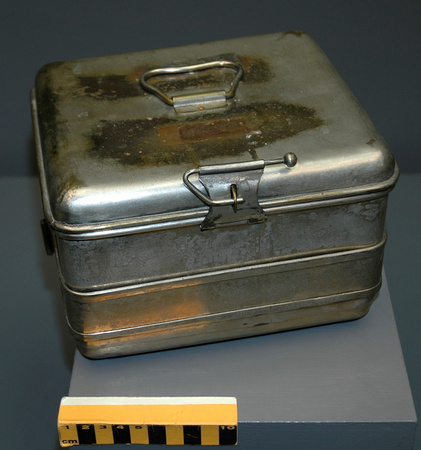
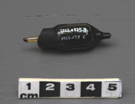
- Markings
- None evident, save UHN catalogue no. "X966.11.44B" printed by hand in white ink.
- Missing
- None.
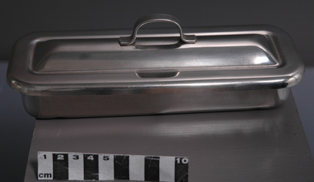
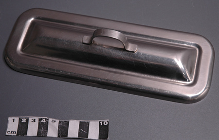
- Finish
- Dull grey metal.
- Decoration
- N/A
CITE THIS OBJECT
If you choose to share our information about this collection object, please cite:
Schering, Blade, circa 1950–1965, Artifact no. 2002.0618, Ingenium – Canada’s Museums of Science and Innovation, http://collection.ingenium.ca/en/id/2002.0618.002/
FEEDBACK
Submit a question or comment about this artifact.
More Like This


2002.0618.002